












Table of contents
- What Is Forensic Radiography?
- Forensic X-ray vs Other Forensic Imaging Methods
- What Is Digital Radiography, and Why Forensic X-ray Runs on It
- What Does a Forensic X-ray Scan Reveal?
- Who Operates Forensic X-ray Systems?
- Types of Forensic X-ray Systems: Stationary, Mobile, and Shielded
- Forensic Imaging at Mass-Fatality and Disaster Sites
- Certifications and Regulatory Compliance
- Choosing the Right FOBOS System for Your Facility
- Why Facilities Adopt Digital Forensic Radiography
- The Future of Forensic Imaging
- Why Forensic Facilities Choose LINEV Systems
- FAQ about X-ray scanners for Forensic Radiography
- Source
Forensic X-ray Systems for Morgues and Forensic Centers
FOCUS: Non-invasive whole-body imaging to detect fractures, foreign objects, and identifying features in human remains before autopsy
APPLICATION: Whole-body screening of remains in morgues, forensic centers, and mobile disaster-response deployments
SOLUTION: Forensic X-ray scanner solutions
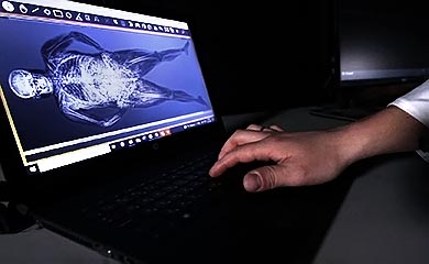
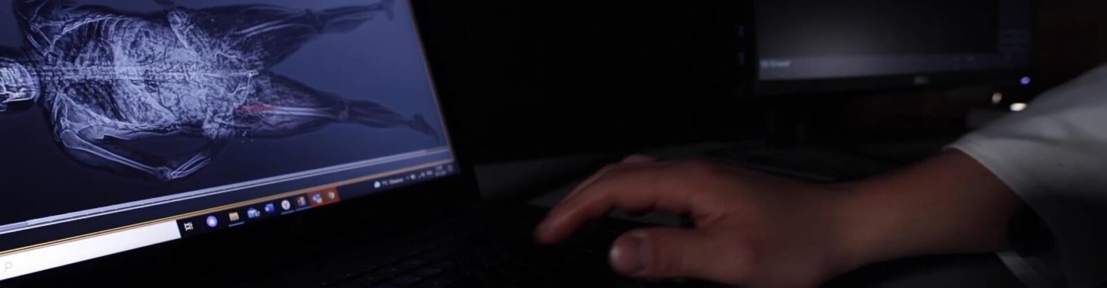
A forensic X-ray system — sometimes called an autopsy imaging system, or referred to under the broader term x-ray forensics — is a digital radiography platform built specifically for post-mortem examination. It captures whole-body, high-resolution images of bones, soft tissue, and foreign objects in under a minute, without altering the body before autopsy. Clinical radiography is built around diagnosing and treating a living patient; forensic radiography answers a different question — what evidence is inside this body, and how do we document it before anyone touches it.
That question has become more urgent as the gap between caseload and capacity keeps widening in the US medicolegal system. A 2019 National Institute of Justice analysis found that the US had only 400–500 full-time forensic pathologists against an estimated need of 1,100–1,200 1. The shortfall hasn’t closed since: as of mid-2026, the National Association of Medical Examiners puts the number of practicing forensic pathologists at roughly 850 nationwide, about 400 short of current demand, according to a forensic pathologist writing in STAT 2. Digital forensic radiography doesn’t fix that shortage, but it changes what a single pathologist can get through in a shift — a full-body scan takes under 30 seconds and tells them, before they pick up a scalpel, whether a case actually needs a full dissection.
What Is Forensic Radiography?
Forensic radiography is the use of X-ray imaging to answer investigative rather than clinical questions: what fractures occurred, is there a foreign object inside the body, does a surgical implant or skeletal feature help establish identity. The physics are the same as in a hospital radiology department; the purpose is not. A clinical X-ray exists to guide treatment. A forensic X-ray exists to build an evidentiary record — and it has to do that without requiring a cooperative, positionable patient, which is precisely why forensic equipment is built differently from the ground up: heavier tables, whole-body coverage in a single pass, and documentation workflows designed for chain-of-custody rather than a discharge summary.
The evidence for using imaging alongside — not instead of — traditional autopsy is well established. A meta-analysis in the International Journal of Legal Medicine, covering 1,757 fracture cases, found postmortem CT reached a pooled sensitivity of 87% and specificity of 97% for fracture detection 3. In cases of sudden cardiac death from coronary artery disease, postmortem CT angiography showed 92% sensitivity and 95% specificity against conventional autopsy as the reference standard4. A broader systematic review of postmortem imaging in trauma cases found that diagnostic performance varies considerably by injury type and anatomical location 5 — which is exactly why radiography is positioned in practice as a triage and documentation layer, not a full replacement for dissection.
It’s worth being direct about that last point with anyone evaluating this equipment: X-ray and CT do not replace autopsy where a definitive cause-of-death finding is legally required. What they do is let a pathologist decide, in under a minute, whether a full dissection is actually necessary for a given case, and leave behind an objective, storable record of internal findings before anyone opens the body — a record a second pathologist, a court, or a researcher can review independently later.
Forensic X-ray vs Other Forensic Imaging Methods
No single imaging modality answers every investigative question, and choosing between them is less about which technology is “better” than about matching the tool to the case and the facility’s operational reality.
| Technology | Main strength | Typical use | Limitation |
| Digital X-ray | Fast, high-resolution projection imaging | Whole-body screening, fracture detection, foreign object localization, and implant identification | Provides less depth information than CT because images are two-dimensional projections |
| Post-mortem CT | Three-dimensional cross-sectional reconstruction of internal anatomy | Complex trauma assessment, gunshot investigations, injury reconstruction, and detailed anatomical analysis | Higher acquisition and operating costs, with greater space and infrastructure requirements |
| MRI | Excellent soft-tissue visualization without ionizing radiation | Specialized forensic investigations, soft-tissue research, and selected post-mortem examinations | Longer scan times, higher operational complexity, and limited availability in forensic facilities |
| 3D surface scanning | High-accuracy documentation of external body surfaces | Identification, documentation of surface injuries, and forensic scene reconstruction | Does not visualize internal anatomy or detect internal injuries |
In most forensic workflows, X-ray is the first imaging step precisely because it’s the fastest way to make that decision. Facilities that adopt it as a triage layer tend to send only the trauma or ballistic cases that genuinely need 3D reconstruction on to CT, instead of running every case through the more expensive modality out of habit.
That triage role also defines FOBOS XR’s economic position relative to CT. Rather than building CT capacity for every post-mortem case, a facility can use FOBOS XR for rapid, routine examination, reserving CT for cases where true three-dimensional reconstruction genuinely adds investigative value. The goal isn’t to reproduce CT’s capabilities — it’s to cover the much broader volume of routine cases at a fraction of the acquisition, operating, and infrastructure cost.
What Is Digital Radiography, and Why Forensic X-ray Runs on It
“Digital X-ray” in the table above isn’t a marketing label — it’s a specific technology, and it’s worth separating from forensic radiography as a practice. In a digital radiography (DR) system, X-rays pass through the body and reach an electronic detector, which converts the signal directly into a digital image rather than exposing film. That one change is behind most of the operational advantages forensic facilities actually notice day to day.
Images become available almost immediately after acquisition instead of waiting on film development — the reason a full-body scan on a system like FOBOS XR takes under 30 seconds rather than the many minutes a film-based process would need. Digital files can be stored securely, sent to another specialist for a second opinion, and adjusted with the same tools radiologists use clinically — contrast, zoom, window/level — without touching the original data.
For a facility processing a steady caseload, that’s not a marginal upgrade. It’s what makes PACS integration, remote consultation between a pathologist and a radiologist, and a permanent, reviewable case archive possible at all. Older film-based or computed radiography (CR) workflows can still technically produce a forensic image, but they can’t match DR’s acquisition speed or plug into a digital case-management workflow the same way — which is why virtually every forensic X-ray system built today, FOBOS included, runs on DR rather than film.
What Does a Forensic X-ray Scan Reveal?
A forensic X-ray identifies skeletal fractures and dislocations, bullet and metal fragments, implanted medical devices and prosthetics, surgical hardware, and structural anomalies useful for identification — all without an incision. That range of findings covers three recurring investigative scenarios.
In trauma cases — road collisions, falls, blunt force, assault — the external appearance of a body often doesn’t reflect the extent of internal injury. Imaging shows fracture location and displacement, and helps establish whether skeletal damage is consistent with the reported circumstances of death.
In ballistic and foreign-object cases central to crime investigation, X-ray shows the exact position of a bullet or fragment before it’s recovered, so investigators have a documented original position rather than relying on memory of where something was before retrieval disturbed it.
In identification cases, orthopedic implants, healed fractures, and surgical hardware can be cross-referenced against medical records — work that becomes especially important in unidentified-remains cases and disaster victim identification (DVI), where visual recognition often isn’t possible at all. INTERPOL’s DVI protocol treats radiographic and dental records as core identification data alongside DNA and fingerprints 6. The operational side of field DVI imaging — working directly at the incident site — is covered in the next section.
Who Operates Forensic X-ray Systems?
Forensic X-ray equipment is typically run by forensic radiographers, radiologic technologists trained in post-mortem imaging protocols — often called a forensic X-ray tech in job postings — or forensic pathology staff working under a medical examiner’s supervision. A forensic radiologist, by contrast, is the physician who interprets the resulting images within the medical-legal context of the case, usually working alongside the pathologist rather than operating the scanner directly. There’s no universal license specific to post-mortem imaging in the US the way there is for clinical radiography — training requirements are set at the facility or state level, usually alongside standard radiation-safety certification. In practice, this means the operator learning curve is shorter than facilities sometimes expect, since most of the workflow (positioning, image acquisition, PACS handling) maps closely onto skills radiologic technologists already have.
Types of Forensic X-ray Systems: Stationary, Mobile, and Shielded
Forensic facilities don’t all face the same constraints, and the industry has settled on three system archetypes because of it. A high-volume medical examiner’s office needs throughput and permanent infrastructure. A disaster response team needs to bring imaging to the bodies, not the other way around. A smaller morgue or a temporary lab often has neither a dedicated X-ray room nor the budget or timeline to build one. LINEV Systems’ FOBOS line covers exactly these three scenarios — full specifications, datasheets, and quote requests are on the Forensic X-ray Scanner Solutions category page.
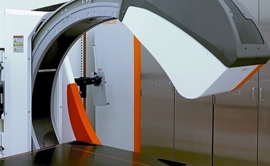
FOBOS XR — Full-body forensic radiography system. The stationary configuration, designed for morgues, coroners’ offices, and forensic centers running a steady, ongoing caseload. It’s built for high-throughput permanent installation, with a 340 kg / 750 lbs capacity table and AP/lateral imaging up to 110°.
FOBOS M — Mobile forensic radiography system. A van-mounted system for disaster victim identification, mass casualty response, and field examinations, for situations where transporting remains to a central facility isn’t practical or possible.
FOBOS XRS — Shielded forensic radiography system. Built-in radiation shielding for facilities that don’t have a dedicated X-ray room — temporary forensic labs, smaller morgues, or sites where constructing a shielded room isn’t feasible on the available timeline or budget.
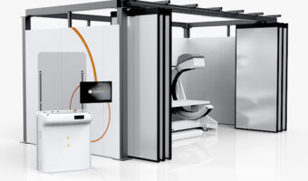


Discover the best forensic solution for premises which are not well X-ray shielded or for temporary forensic laboratories
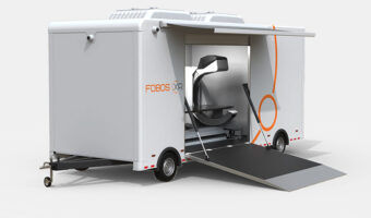
The mobile forensic solution consisting of FOBOS system mounted in van
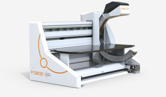

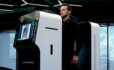
The perfect forensic X-ray imaging solution for morgues, coroners and forensic centers
Forensic Imaging at Mass-Fatality and Disaster Sites
Mass-fatality incidents create a different problem from routine morgue work: imaging capacity may be required where the remains are located, while large numbers of cases have to be examined and documented within a limited period of time. Transporting every case to a hospital or central forensic imaging facility can introduce delays and additional handling at exactly the point where investigators need information quickly.
A mobile forensic radiography system brings the imaging process directly to the disaster-response site. Whole-body digital X-ray examination can give investigators and forensic teams an immediate view of skeletal trauma, foreign objects, medical implants, and other internal findings that may assist with preliminary assessment, identification, and case prioritization. The examination can be performed before extensive handling or invasive examination of the remains, creating a digital record that pathologists, radiologists, and identification teams can review afterward.
For disaster victim identification, this combination of mobility and speed is particularly important. FOBOS M allows radiographic information to be acquired close to the incident location and incorporated into the DVI workflow immediately, rather than after remains reach a permanent facility. In situations where remains arrive enclosed in protective transport packaging or body bags, radiography can also provide an initial non-invasive view before further examination and handling, subject to the applicable examination protocol and the radiographic properties of the packaging.
Certifications and Regulatory Compliance
For whoever signs off on a capital request, this is usually the first thing checked, and it’s worth having answered before the question gets asked. Diagnostic X-ray systems and their major components sold in the US fall under the federal performance standard at 21 CFR 1020.30, which covers radiation output, beam limitation, and manufacturer certification and assembly reporting 7. LINEV Systems’ quality management system is ISO 9001 certified, covering design, manufacturing, and service processes across the FOBOS line. Installation of a stationary unit still needs to account for state-level shielding and radiation-protection requirements — this is exactly the gap the shielded FOBOS XRS configuration is designed to close for facilities that don’t already have that infrastructure in place. Keeping FDA and ISO documentation ready as attachments to a purchase request, rather than just referenced by name, tends to shorten the review cycle with procurement and safety committees considerably.
Choosing the Right FOBOS System for Your Facility
The right system depends less on which one has the most features and more on which operational profile matches your facility.
FOBOS XR fits an office running a steady, year-round caseload out of a fixed location that wants imaging folded into an existing PACS-based workflow: permanent installation, sub-30-second full-body scans, and a table designed around the body sizes and weights a busy morgue actually sees rather than an idealized clinical patient.
Disaster response and mass casualty work changes the deciding factor entirely — it stops being about image quality and becomes about whether the system can get to the bodies at all, at sites where remains can’t be moved to a central facility. FOBOS M is built around that constraint: a van-mounted, field-deployable configuration that keeps the same digital workflow and PACS integration as the stationary system, just portable.
For facilities without a dedicated X-ray room — a smaller morgue, a temporary lab, a site where a shielding construction project isn’t realistic on the timeline — the installation question usually decides the purchase before the imaging specs do. FOBOS XRS solves that by building the shielding into the system itself, so deployment doesn’t wait on a construction budget.
Beyond matching the configuration to your operational profile, a few practical questions are worth putting in front of any vendor, LINEV included: how the system’s PACS output integrates with your existing case management software rather than creating a second, disconnected archive; what the manufacturer’s service response time and parts availability actually look like, since downtime on a single forensic imaging system stalls every case behind it; and what training and onboarding time to budget for staff who haven’t operated whole-body forensic equipment before.
Why Facilities Adopt Digital Forensic Radiography
The case for digital radiography in a forensic setting isn’t a single advantage — it’s that several operational pressures point the same direction at once. Evidence preservation is the clearest one: internal findings get documented before any invasive procedure has a chance to change them, which matters directly in cases where cultural, religious, or legal considerations limit how much dissection is appropriate. Triage is the second, and arguably the one with the biggest day-to-day impact on a backlogged office — a scan that takes under a minute tells a pathologist which cases genuinely need a full autopsy and which don’t, instead of defaulting every case to the most invasive and time-consuming option available.
There’s also a collaboration effect that’s easy to underestimate until an office actually has it: once a case exists as a digital image set, a pathologist, a radiologist, and an investigator can each review the same findings independently, without needing repeated physical access to the remains, and without waiting on each other’s schedules. And because the record is permanent and storable, it holds up for exactly the situations where a single physical examination isn’t enough — a case reopened years later, a second opinion requested by a defense attorney, or a training file used to teach the next generation of forensic pathologists.
The Future of Forensic Imaging
Forensic imaging is moving toward tighter integration with case management systems and, increasingly, AI-assisted image review. AI tools are being researched for fracture-detection assistance, pattern recognition, and workflow triage — but interpreting findings within the legal and investigative context of a case remains a qualified specialist’s responsibility, not something an algorithm decides. Disaster victim identification is a good example of where standardization is already mandatory rather than optional: INTERPOL’s DVI Guide, most recently updated in 2023, sets the international protocol for documenting ante-mortem and post-mortem data across jurisdictions 6, and imaging records are a standard part of that documentation chain — which is exactly the kind of workflow a mobile, field-deployable system like FOBOS M is built to plug into.
Why Forensic Facilities Choose LINEV Systems
FOBOS systems are built specifically around forensic workflows rather than adapted from clinical radiography equipment — the weight capacity, whole-body single-pass coverage, and PACS integration described throughout this page reflect design choices made for morgues and forensic centers, not retrofits of hospital equipment. That focus extends past active casework: university forensic pathology programs and research groups use the same systems to study trauma patterns, train the next generation of examiners, and compare imaging techniques, without repeated physical examination of remains and with a level of image consistency that’s hard to reproduce on adapted clinical equipment.
Combined with FDA-compliant design, ISO 9001–certified manufacturing, and coverage of the three deployment scenarios forensic organizations actually face — permanent installation, field mobility, and shielded retrofit — it’s a narrower, more purpose-built choice than adapting general-purpose imaging equipment to forensic use.
If you’re evaluating a forensic X-ray system for your facility, request a FOBOS datasheet and quote to compare configurations against your caseload, infrastructure, and budget directly with our team.
FAQ on Forensic X-ray Systems
No. Imaging is a triage and documentation tool, not a legal substitute for autopsy where a jurisdiction requires a definitive cause-of-death finding through dissection. Its role is deciding which cases need a full autopsy and which don’t, and preserving evidence either way.
Four parts working together: a high-output X-ray generator, a digital detector that converts the X-ray signal into an image, a positioning table or cart built for whole-body imaging rather than a single limb, and PACS-integrated acquisition software for storing and retrieving cases. The specific combination — generator power, detector resolution, cart capacity and adjustability — is what actually differentiates one system from another, more than any single spec on its own.
Forensic radiography is the technique — acquiring the X-ray image. Forensic radiology is the medical specialty of interpreting those images within a legal and investigative context, typically practiced by a radiologist working alongside the forensic pathologist rather than operating the equipment.
Digital forensic images are routinely used as supporting evidence in criminal and civil proceedings, similar to photographs or other documented physical findings, though admissibility standards and chain-of-custody requirements vary by jurisdiction and should be confirmed with local legal counsel rather than assumed.
Operator exposure is managed through standard radiation-protection practice — shielded operator positions, exposure controls, and, where the facility lacks a shielded room, built-in shielding like that in the FOBOS XRS. Systems sold in the US are also subject to FDA performance standards under 21 CFR 1020.30 governing radiation output 7.
Not necessarily. A permanent installation like FOBOS XR is typically installed in a shielded room built for the purpose, but shielded configurations such as FOBOS XRS are designed specifically for facilities without one, removing the construction project from the purchase timeline.
Yes — that’s the primary use case for van-mounted systems like FOBOS M, which bring whole-body imaging and PACS documentation to a disaster site instead of requiring remains to be transported to a central facility first.
Installation and any required shielding work, staff training time, PACS integration with existing case management software, and ongoing service and maintenance costs are the recurring line items that affect total cost of ownership more than the sticker price does.
Source
1. National Academies Press, Strengthening the U.S. Medicolegal Death Investigation System, National Institute of Justice data analysis, 2019.
2. McDonald, G., “The shortage of forensic pathologists is hurting justice, public health, and families”, STAT First Opinion, citing National Association of Medical Examiners (NAME) data, July 2026.
3. International Journal of Legal Medicine, “Comparison of the diagnostic efficacy of postmortem computed tomography and traditional autopsy in fracture detection: a meta-analysis”, 2025.
4. La radiologia medica, “Postmortem computed tomography angiography (PMCTA) and traditional autopsy in cases of sudden cardiac death due to coronary artery disease: a systematic review and meta-analysis”, 2018.
5. International Journal of Legal Medicine, “Postmortem imaging findings and cause of death determination compared with autopsy: a systematic review of diagnostic test accuracy and meta-analysis”, 2019.
6. INTERPOL, Disaster Victim Identification Guide, November 2023.
7. U.S. Food and Drug Administration, 21 CFR 1020.30: Diagnostic X-ray Systems and Their Major Components.